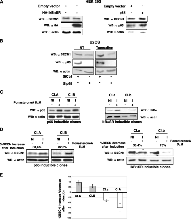
FIG. 4.
BECN1 protein level regulation by p65. (A) Lysates from HEK293 cells overexpressing hemagglutinin-tagged human IκBαSR, p65, or an empty vector were subjected to sodium dodecyl sulfate-polyacrylamide gel electrophoresis and subsequently analyzed by immunoblotting for BECN1 protein expression. (B) BECN1 protein levels were evaluated by immunoblotting in untreated and tamoxifen (10 μM, 5 h)-treated U2OS cells following p65 depletion. Ctrl, control. (C) Generation of U2OS cell lines inducible for either p65 or IκBSR. Inducible cell lines were obtained as described in Materials and Methods. Two clones for each kind of inducible U2OS cells were tested. Treatment with 5 μM ponasterone A (I) for 16 h significantly increased the expression of both p65 and IκBαSR in the selected clones with respect to that in uninduced (NI) cells. (D, left) U2OS clones A and B, inducible for p65, were treated with 5 μM ponasterone A (I) or left untreated (NI) for 6 h. Cell lysates were subjected to Western blot (WB) analysis for monitoring of BECN1 levels. (D, right) Induction of IκBSR clones a and b was preformed, and BECN1 levels were assessed by immunoblotting. (E) Quantification of the bands was performed with the ImageJ tool. The graph is representative of three independent experiments performed with each of the four clones. Percentages of BECN1 protein increase or decrease following ponasterone A treatment are reported.

