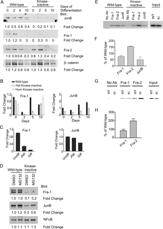
FIG. 8.
JNK inhibition alters AP-1 composition in TS cells. (A) Jun B and Fra-1 protein levels are significantly reduced in kinase-inactive TS cells. Results from Western blot analysis of nuclear lysates from wild-type and heterozygote kinase-inactive TS cells differentiated for the indicated times are shown. Blots were probed with antibodies against JunB, Fra-1, Fra-2, and β-catenin. Changes are relative to undifferentiated wild-type TS cells (time zero). (B) Fra-1 and JunB mRNA levels are significantly reduced in kinase-inactive TS cells. Fra-1 and JunB mRNA levels were measured by quantitative RT-PCR of TS cells differentiated for the indicated number of days. Data are the means ± ranges of two experiments. Data were normalized to wild-type undifferentiated levels. (C) Inhibition of JNK with SP600125 in undifferentiated TS cells induces the loss of Fra-1 message. Wild-type TS cells were treated for 2 days under undifferentiating conditions with DMSO (Undiff), 50 μM SP600125 (JNK inhibitor), or under differentiating conditions with DMSO (Diff). Fra-1 and JunB mRNA levels were measured by quantitative RT-PCR. Data are the means ± ranges of between two and four independent experiments. Data were normalized to wild-type undifferentiated levels. (D) Treatment with the proteasome inhibitor MG132 does not restore Fra-1 levels in kinase-inactive TS cells. Undifferentiated TS cells were treated with DMSO or MG132 for 5 hours and analyzed as described for panel A. (E) Alteration in AP-1 family member binding to the third intron of Gcm1 in undifferentiated MEKK4WT/K1361R cells. ChIP analysis was performed using antibodies to Fra-1, Fra-2, and JunB using samples obtained from undifferentiated MEKK4WT and MEKK4WT/K1361R TS cells. PCR was performed to a region containing an AP-1 consensus sequence (TGA G TCA) in the third intron of Gcm1. KI, kinase inactive. (F) Quantitation of ChIP analysis of Gcm1. Data shown are the means ± ranges of between two and three experiments. Bands were quantitated by densitometry using ImageJ and data were normalized to wild-type levels. (G) Alteration of AP-1 family member binding to the MMP2 promoter in undifferentiated MEKK4K1361R cells. ChIP analysis was performed with antibodies to Fra-1 and Fra-2 using samples obtained from undifferentiated MEKK4WT and MEKK4K1361R TS cells. PCR was performed to a region in the MMP2 promoter (−2723 to −2344) containing an AP-1 consensus sequence (TGA C TCA). (H) Quantitation of ChIP analysis results with the MMP2 promoter. Data shown are the means ± standard errors of the means of three experiments.

