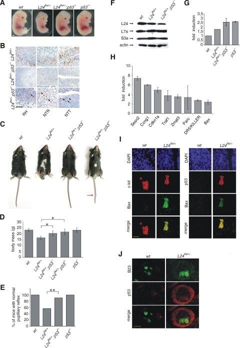
FIG. 6.
p53 inactivation suppresses the phenotype of Rpl24Bst/+ mice. (A) Morphology of representative (i) wt, (ii) Rpl24Bst/+, and (iii) Rpl24Bst/+; p53−/− embryos at E13.5. (B) Apoptosis in roofs of hindbrains (RH), neural tubes in the trunks (NTR), and neural tubes in the tails (NTT) of E10.5 (i) Rpl24Bst/+, (ii) Rpl24Bst/+; p53−/−, and (iii) Rpl24Bst/+; p53+/− embryos. Apoptotic cells are indicated by arrows. (C) The phenotype of adult Rpl24Bst/+ mice is suppressed by p53 inactivation. Shown are photographs of representative (i) wt, (ii) Rpl24Bst/+, and (iii) Rpl24Bst/+; p53−/− mice (n = 16) and Rpl24Bst/+; p53+/− mice (n = 54) at 6 weeks of age. A kink in the tail is indicated by the red arrow. (D) Mean body masses of wt (n = 18), Rpl24Bst/+ (n = 10), Rpl24Bst/+; p53−/− (n = 14), Rpl24Bst/+; p53+/− (n = 23), and p53−/− (n = 17) male mice at 6 weeks of age. We analyzed males because only two female Rpl24Bst/+; p53−/− mice were recovered. *, P < 0.05 (Mann-Whitney test). (E) Percentage of wt (n = 32), Rpl24Bst/+ (n = 30), Rpl24Bst/+; p53+/− (n = 32), and p53+/− (n = 32) mice with the normal pupillary reflex. **, P = 0.006 (chi-square test). Equal numbers of males and females were analyzed. (F) The levels of Rpl24 protein in lysates of E10.5 wt, Rpl24Bst/+, and Rpl24Bst/+; p53−/− embryos were determined by immunoblotting with Rpl24 rabbit polyclonal antibody. Membranes were reprobed with antibodies to Rpl7a, Rps3a, and actin. (G) RNA was isolated from E10.5 embryos of the indicated genotypes. A real-time PCR using 47S rRNA probes was performed in triplicate. The SD was calculated on the basis of three independent experiments. (H) Real time RT-PCR analyses of 84 selected p53 target genes. Eight genes were upregulated more than twofold in E10.5 Rpl24Bst/+ embryos compared to wt embryos. The SD was calculated on the basis of three independent experiments. (I) Transversal sections of the trunk neural tube of E10.5 wt and Rpl24Bst/+ embryos were costained with a c-kit and anti-Bax (left panel) or anti-p53 and anti-Bax (right panel) antibodies and analyzed by fluorescence microscopy. Nuclei are stained with DAPI (blue). (J) Transversal sections of the trunk neural tube of E10.5 wt and RPL24Bst/+ embryos were stained with p53 antibody (red) and B23 (green) and analyzed by confocal laser scanning microscope. Error bars in panels D, G, and H indicate the SD. Scale bars: 1 mm (A), 100 μm (B), 50 μm (I), and 5 μm (J).

