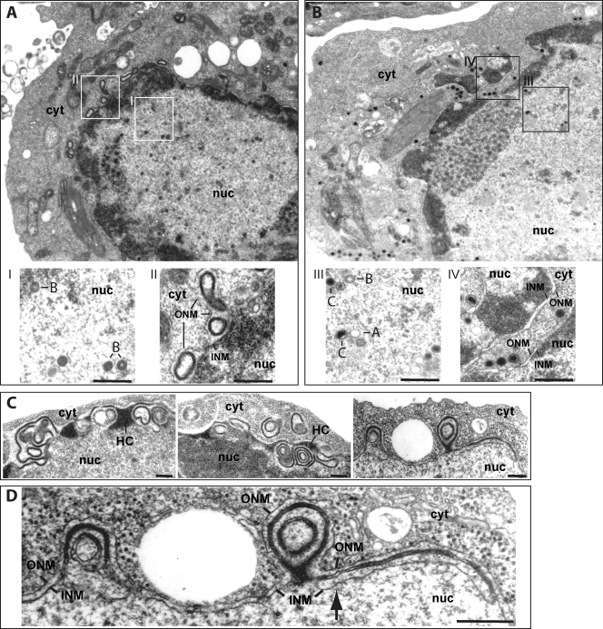
FIG. 3.
Electron micrographs of induced 293/ΔBGLF5 (A, C, and D) and 293/ΔBGLF5-C cells (B). (A) Overview of a replicating 293/ΔBGLF5 cell at low (×8,000) magnification. The nucleus contains numerous immature B capsids with scaffolding structures but only rare mature C-capsids carrying electron-dense DNA (inset I). No viral structures could be detected in the cytoplasm. The nuclear membrane was highly irregular in shape and displayed electron-dense membrane foldings and/or duplications within the perinuclear space resulting in the formation of nuclear pockets, some of which were deeply located in the cytoplasm (inset II). (B) Complementation of 293/ΔBGLF5 cells corrected these abnormalities; all three types of nucleocapsids (inset III) and mature capsids were visible in the cytoplasm, and the nuclear membrane displayed normal morphological features (inset IV). (C and D) Several micrographs provide a more detailed view of the nuclear membrane from induced 293/ΔBGLF5 cells at lower and high magnifications. The nuclear membrane appears thickened and by places connected to heterochromatin (panel C, left and middle images). The cellular material accumulated in the nuclear membrane has a similar density as heterochromatin. The remaining pictures (panel C, right image, and panel D) exemplify what are suspected to be earlier events; the nuclear membrane appears duplicated with four stacked membrane layers visible (arrow). The electron-dense material appears to progressively fill in the space between nuclear membranes to form multiple projections of nuclear envelope that produce an appearance of concentric nuclear pockets on these two-dimensional sections. Note that some nuclear pockets enclose a profile of cytoplasm (panel B, middle image). INM, inner nuclear membrane; ONM, outer nuclear membrane; nuc, nucleus; cyt, cytoplasm; HC, heterochromatin; A, B, and C, A-, B-, and C-type capsids. Bar, 0.2 μm.

