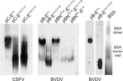
FIG. 3.
Blue native PAGE of Erns proteins from different pestiviruses. Proteins were expressed in BHK-21 cells and lysed with a lysis buffer containing 1% digitonin. Proteins were separated through blue native PAGE with a histidine-containing buffer system (40) and blotted onto nitrocellulose membranes. The transfected plasmids are indicated above the lanes (see the legend to Fig. 2 for designations). Detection was carried out with an Erns-specific serum. On the right, the results of a control experiment are shown, with the left two lanes containing proteins expressed from pB-Erns and pB-Erns-C171R (equivalent to lanes 1 and 2 of the middle panel) and the right lane showing a Coomassie blue-stained BSA marker. The monomeric and dimeric bands of the marker protein are visible, corresponding to ca. 66 and 132 kDa, respectively.

