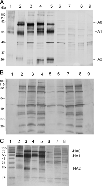
FIG. 4.
Western blots of cell lysates probed for influenza virus HA expression (A) or vaccinia virus antigen expression (B). Lane 1, formalin-inactivated purified influenza virus. Lane 2, replicating virus rVV-HA-VN (VN1203) grown in Vero, MOI of 0.01. Lane 3, replicating virus rVV-HA5 (VNHY/04), Vero, MOI of 0.01. Lane 4, defective virus dVV-HA5 (VNHY/04), cVero22, MOI of 0.01. Lane 5, dVV-HA5 (VNHY/04), Vero, MOI of 5; lane 6, negative control dVV-L, Vero, MOI of 5.0; lane 7, dVV-L, cVero 22, MOI of 0.01; lane 8, Lister wt, MOI of 0.01; lane 9, negative control, uninfected Vero cell lysate. (C) HA expression in wt Vero cells infected with the different vectors at a constant MOI of 5. Lane 1, size markers; lane 2, positive control, 0.5 μg of the formalin-inactivated influenza vaccine; lane 3, replicating virus rVV-HA-VN; lane 4, replicating virus rVV-HA5; lane 5, defective virus dVV-HA5; lane 6, empty vector dVV-L; lane 7, empty vector VV-L; lane 8, uninfected Vero cell lysate.

