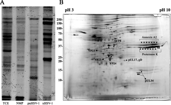
FIG. 5.
Analysis of primary enveloped virion protein profile. (A) Coomassie-stained 10% SDS-PAGE gel shows the protein profile of TCE, NMP, pnHSV-1, and xHSV-1. (B) Silver-stained 2D gel displays the pnHSV-1 fraction protein profile. Arrowheads denote the spots picked that corresponded with the proteins identified and listed in Table 1.

