FIG. 9.
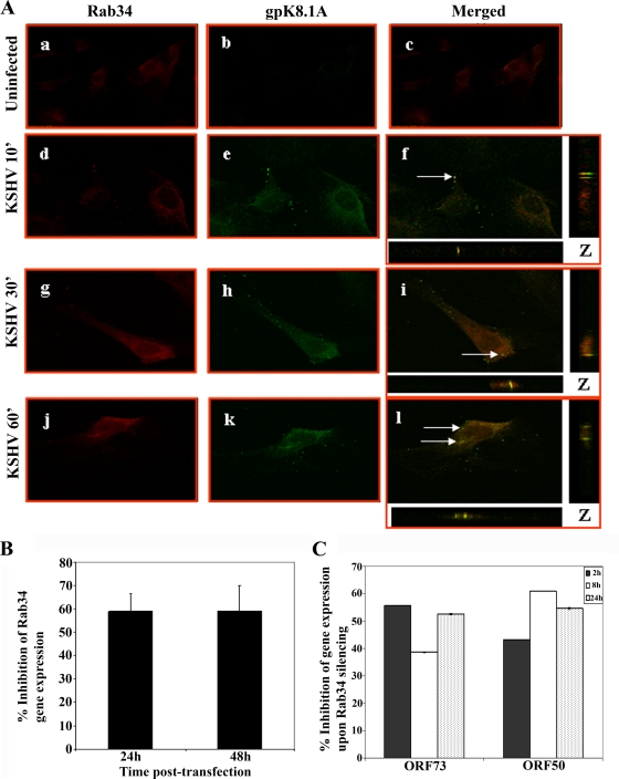
KSHV trafficking in HMVEC-d cells and association with Rab34. (A) HMVEC-d cells grown in eight-chamber slides were infected with KSHV at an MOI of 10 for different times, washed, fixed in 4% paraformaldehyde, permeabilized, and stained with goat anti-Rab34 antibodies and with anti-gpK8.1A mAb for 1 h at room temperature. The cells were washed and incubated with anti-mouse Alexa Fluor 488 (green) and anti-rabbit Alexa 594 Fluor (red) antibodies, washed, mounted, and visualized with a confocal laser scanning microscope, and the data analyzed by using Olympus Fluoview software. Arrows indicate areas of colocalization of virion particles with Rab34. Magnification, ×40. ', min. (B) Histogram representing percent inhibition of Rab34 gene expression in HMVEC-d cells after 24 h and 48 h of transfection with si-Rab34 and si-C as described in Materials and Methods. Percent inhibition was calculated using Rab34 expression in si-C-transfected HMVEC-d cells as 100%. Each bar represents the average ± standard deviation of the results of three independent experiments. (C) Histogram representing the percent inhibition of ORF50 and ORF73 gene expression upon Rab34 silencing in HMVEC-d cells. The percent inhibition was calculated by considering levels of ORF50 or ORF73 in si-C-transfected cells infected with KSHV to be 100%. Each bar represents the average ± standard deviation of the results of three independent experiments.

