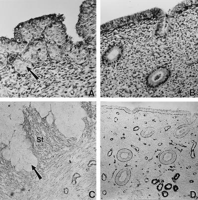
Figure 2.
Morphology (A and B) and immunolocalization of αSMA (C and D) in cycling baboons treated with hCG. Note the distinct epithelial plaques that are associated with the luminal epithelium (arrow; A) and the localization of αSMA in stromal fibroblasts below the plaques (St; C) in hCG-treated animals. The controls treated with heat-inactivated CG showed no response in either of the two cell types (B and D). [Final magnification ×77 (A and B) and ×45 (C and D).]

