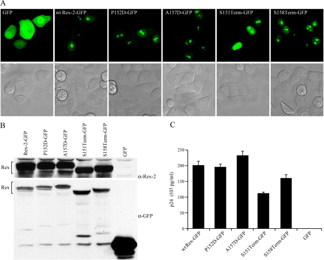
FIG. 2.
Subcellular localization of Rex-2 mutants. HeLa-Tat cells were transfected with 1 μg of various Rex-2-EGFP plasmids or the EGFP-N3 negative control as indicated using Lipofectamine Plus (Invitrogen, Carlsbad, CA). (A) For EGFP detection, cells were plated and visualized using a Zeiss LSM 510 microscope (GFP and the light field are shown). (B) Expression of Rex-2-EGFP fusion proteins was detected by Western blot analysis using anti-Rex-2 antisera or anti-EGFP antibody. α, anti. (C) The functional activities of Rex-2-EGFP fusion proteins were determined by using an HIV p24 Gag reporter assay. The values represent actual p24 Gag production from a representative experiment performed in triplicate. Error bars indicate standard deviations.

