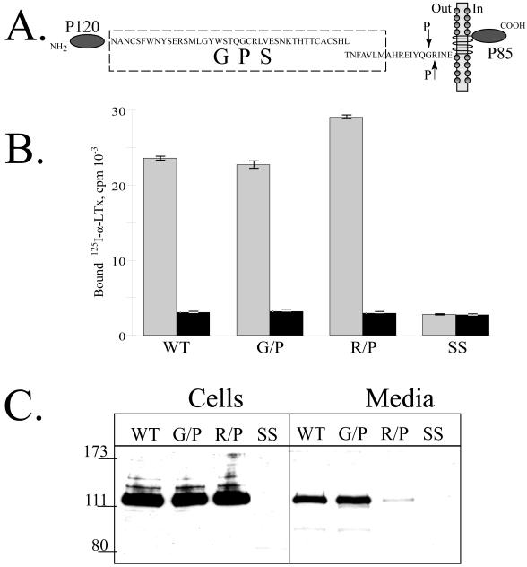
Fig. 7. Cell surface expression of CIRL mutated at the second cleavage site.
A. Schematic description of the CIRL constructs with single residue mutations (G852/P and R853/P, indicated by arrows) at the second cleavage site. B. Cell surface expression of the second cleavage site CIRL mutants. Intact COS cells transfected with either wild-type CIRL or its mutants (G852/P and R853/P) were assayed for binding of 125I-α-latrotoxin either in the presence (black bars) or absence of excess non-labeled α-latrotoxin. Measurements were performed in triplicates. C. Secretion of the soluble forms of CIRL mutants. One ml of conditioned media of the same cells as in B was precipitated with 10 μl of α-latrotoxin-agarose and the eluates were analyzed by Western blotting with anti-p120 antibody. SS, salmon sperm DNA transfected cells. The picture shown is representative of three independent transfection, precipitation and blotting experiments that produced essentially similar results.

