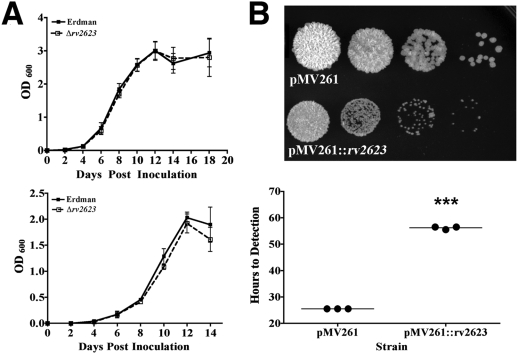
Figure 2. In vitro characterization of Δrv2623.
(A) Growth curves of cultures inoculated (106 CFU/ml) into 7H9+10% OADC+0.05% Tween 80 (top) and in minimal Sauton's media (bottom); Erdman (closed boxes, solid line) and Δrv2623 (open boxes, dashed line) cultures. Error bars represent the standard error of the means; each curve is a combination of at least three independent experiments. (B) Overexpression of Rv2623 in M. smegmatis. The top panel represents serial dilutions (1∶10) of the empty vector pMV261-containing negative control strain and Rv2623-overexpressing strain harboring pMV261::rv2623. Diluted stationary phase M. smegmatis culture was spotted (5 µl) onto solid 7H10 media supplemented with 10% OADC and kanamycin (40 µg/mL). Photographs were taken after three days incubation at 37°C. Growth of corresponding strains in liquid medium was assessed based on the time to detection determined using a BD BACTEC 9000MB system (bottom). The various strains were inoculated at 104 CFU/ml in triplicates. Data shown are representative of several independent experiments. ***p<0.001.

