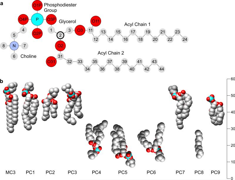
Figure 1.
The structure of dimyristoylphosphatidylcholine (DMPC). (a) Schematic diagram of DMPC with atoms annotated following the nomenclature used in the PBD file of ovine junctional AQP0 (PDB 2B6O) [8]. The chiral C2 carbon of the glycerol backbone is shown bold. (b) Sphere models of the atomic structure of the B form of DMPC (MC3) [9] and the nine resolved lipids in the AQP0 structure. The lipids are positioned at their appropriate positions within the membrane but rotated to allow a comparison with the crystal structure.

