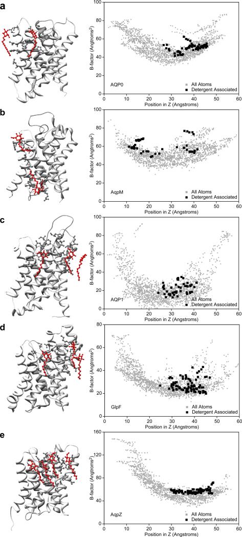
Figure 5.
Detergent-lipid interactions in different aquaporin crystal structures. (a) Bovine AQP0, (b) AqpM from Methanobacterium thermoautotrophicum, (c) bovine AQP1, (d) GlpF from Escherichia coli, and (e) AqpZ from Escherichia coli. The left panels show the crystal structures as ribbon diagrams and the residues within 5 Å of an ordered detergent molecule (red) as ball-and-stick representations. The right panels show plots of the temperature factors of all the atoms in the structures against their position in the direction normal to the membrane plane. The temperature factors of the detergent-contacting atoms are shown in black and those of the other atoms in grey.

