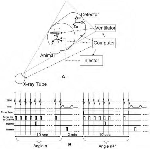
Fig.2.
DSA and TDSA require integration of the X-ray imaging chain ( x-ray tube & detector) with the biological pulse sequencer (gating). (A) shows the schematics of the system during sampling and includes two reference systems i.e. one for the rotating animal or object (x, y, z) and one for the static imaging system (xo, yo, zo). The Biological Pulse Sequence (B) shows suspension of ventilation and image capture on every heartbeat at the QRS complex before and after a single contrast injection. A complete DSA sequence at angle n is acquired in 10 secs of suspended respiration (end expiration). The animal is rotated to the next angle n+1 and ventilated for 2 mins before another DSA sequence is repeated.

