Figure 2.

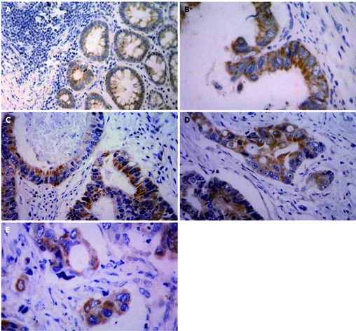
TSPAN1 expression in normal tissues (A), colon cancer tissues (B, C), rectal cancer tissues (D, E). Paraffin section of human colorectal carcinoma tissues was stained with anti-TSPAN1 polyclonal antibody by immunohistochemistry. A: TSPAN1 weakly expressed in the cytoplasm. (× 100). B, C: TSPAN1 was located in the cytoplasm with yellow granulation. (× 200). D, E: Cancer nest showed positive TSPAN1 expression and vascular invasion. (× 200).
