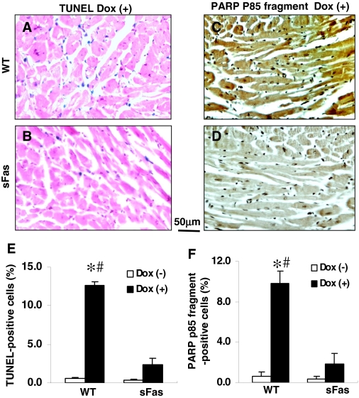
Fig. 5.
Dox-induced cell death in the myocardium is attenuated by expression of sFas. A and B, representative photomicrographs of TUNEL-stained sections obtained from Dox-treated WT mice and Dox-treated sFas mice. Blue staining, TUNEL-positive cells. C and D, representative photomicrographs of immunohistochemical staining demonstrated a strong immunoreactivity for PARP p85 fragment in the myocardial sections from Dox-treated WT mice compared with very little immunoreactivity in the myocardium of Dox-treated sFas mice. Immunoreactivity was visualized with diaminobenzidine (brown). E and F, histograms showing a marked decrease in TUNEL-positive cells and PARP p85 fragment immunoreactivity, respectively, in the myocardium of Dox-treated sFas mice compared with Dox-treated WT mice. n = 5 per group; *, p < 0.01 versus saline-treated WT and sFas mice; #, p < 0.05 versus Dox-treated sFas mice.

