Fig. 8.
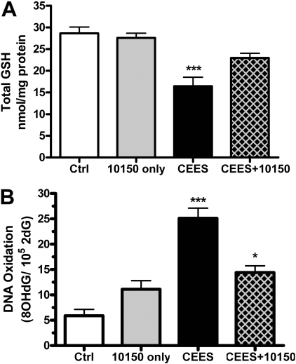
The effects of CEES on markers of cellular oxidative stress and prevention by AEOL 10150 in 16HBE cells. Cells exposed to 900 μM CEES for 12 h had decreased total cellular GSH levels (A), and AEOL 10150 (50 μM) rescued this decrease when treated 1 h after CEES exposure. Total GSH levels were normalized to the amount of protein and expressed as nanomoles of GSH per milligram of protein. CEES also increased the levels of the DNA oxidation marker 8OHdG (B), and AEOL 10150 (50 μM) post-CEES treatment decreased the levels of DNA oxidation. Data expressed as a ratio of 8OHdG per 105 2dG. Data presented as mean ± S.E.M., n = 4 to 8; *, p < 0.05; ***, p < 0.001 compared with control levels. Two-way ANOVA of AEOL 10150, p = 0.1444; CEES, p = 0.0001; interaction, p = 0.0481 (A); and AEOL 10150, p = 0.1394; CEES, p < 0.0001; interaction, p = 0.0004 (B).

