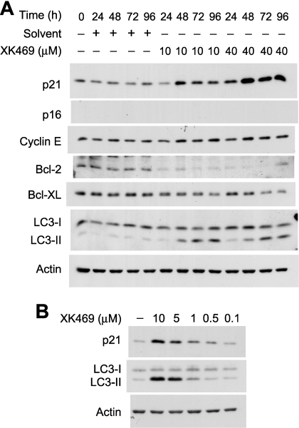
Fig. 9.
Western blot analyses of autophagy, cell cycle, and Bcl-2 proteins. Melan-A cultures were treated with 10 or 40 μM XK469 for different lengths of time (A) or with 0.1 to 10 μM XK469 for 2 days (B) before being harvested for subsequent Western blot analyses. Analyses are of 25 μg protein/lane. Similar results were obtained in a second experiment.

