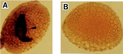
Figure 2.
In situ hybridization of LGC1 mRNA to whole-mounted lily pollen. Dark staining in the generative cell (arrowhead) represents hybridization signal detected by using an alkaline phosphatase conjugated anti-DIG antibody. The outer wall of pollen, exine, appears as a sculptured pattern. (A) Pollen probed with a DIG-UTP-labeled LGC1 antisense riboprobe. (B) Control pollen probed with a sense riboprobe.

