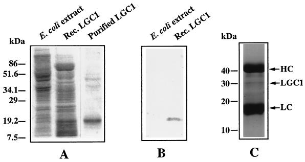
Figure 4.
Immunoblot analysis of LGC1 protein. (A) Coomassie blue-stained SDS/PAGE gel showing the recombinant LGC1 protein. (B) Immunoblot probed with anti-LGC1 antiserum showing specific binding of antibody to LGC1 protein. (C) LGC1 protein immunoprecipitated from total protein extract of generative cells. Two strong bands represent the heavy chain (HC) and light chain (LC) of Ig, respectively.

