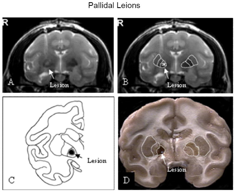
Figure 1.

Location of pallidal lesions. Pallidal lesions were shown in coronal post-operative T2-weighted MR images (A & B). A line drawing was used to illustrate the effective lesion and surrounding edema (C). The pallidal lesion was shown in 4-mm thick coronal section through the globus pallidus (D).
