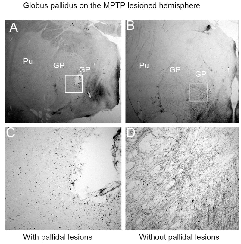
Figure 2.

TH staining in coronal section through the GP on the side ipsilateral to MPTP administration. A typical location of the pallidal lesion was demonstrated in panel A, which was primarily in the internal segment of the GP. The same structure without the lesion from another animal is shown in panel B. High magnification of regions surrounding the lesion revealed a massive loss of the TH positive fiber in the lesioned internal segment of the GP(C), while abundant TH positive fibers are visible in the same areas without a pallidal lesion(D).
