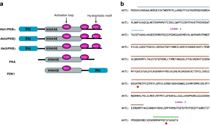
Fig. 1.
PKB/Akt primary structure. a Structural alignment of selected members of the AGC superfamily of protein kinases: the three protein kinase B (PKB/Akt) isoforms, phosphoinositides-dependent protein kinase 1 (PDK1) and protein kinase A (PKA). A high homology exists at the level of the kinase domains (grey). The residues highlighted in pink represent the phosphorylation sites important for the activity of the kinases. PH domains are represented in blue. b Amino acid sequence of PKBα/Akt1. The isolated sections of the protein that have been crystallised are highlighted in blue (PH domain), orange (kinase domain) and green (hydrophobic motif). The red stars indicate the residues Thr 308 and Ser 473. The domains are separated by linker regions that have not been crystallised. The linker 1 is between the PH domain and the kinase domain and the linker 2 is between the kinase domain and the C terminus

