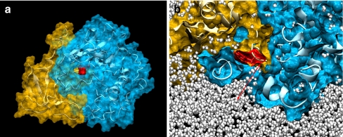
Fig. 4.
PKBα PH domain creates a cavity in the kinase domain. a Structure of the PKBα PH domain (gold) and reconstructed kinase domain (blue) in complex (PKBα “PH-in” conformer) after short dynamic runs and minimisation in water and physiological salt concentration. The secondary structure of the kinase domain is represented as a white ribbon. The PH domain residue Trp 80 (red) is visible through an open cavity in the kinase domain. b Vertical cross section of the dynamic structure shown in a at the level of the PH-induced cavity. The distance between the Trp 80 residue in red and the protein surface is about 8–8.5 Å (red arrow). The cavity is filled with water molecules shown as white pearls

