
Fig. 5.
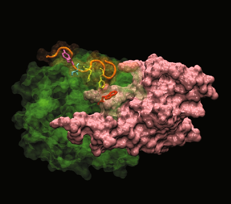
The C-terminal hydrophobic motif of PKBα in the “PH-in” conformer is positioned at the apex of the cavity PKBα C-terminal hydrophobic motif HM (orange) interacting with PKBα kinase domain (green) in the “PH-in” conformer. The PH domain is in pink and the PH domain residue Trp 80 is in red. The model shows that two phenyalanines (in yellow) from the sequence F469XXF472S of HM are found positioned right above the PH-induced cavity in PKBα kinase domain. Ser 473 is represented in cyan and Tyr 474 in magenta