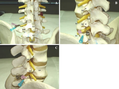
Fig. 2.
Model images of needle placement for a left L4 TFESI. The brown needle illustrates the subpedicular approach, the blue needle shows the retroneural approach, and the pink needle depicts the retrodiscal approach. a AP view. The subpedicular and retroneural approaches have the same target point at the “6 o’clock” position of the L4 pedicle (P). The retrodiscal approach target is lateral to the L5 superior articular process (SAP). b Left oblique view. The subpedicular and retroneural approach have the same target point at the “6 o’clock” position of the L4 pedicle (P). The retrodiscal approach target is lateral to the L5 SAP (overlapped by the brown needle). TP = transverse process. c Lateral view. The subpedicular approach target area lies on the back of the L4 vertebral body. The retroneural approach target area lies more dorsal in the L4–L5 foramen underneath the L4 pedicle (P). The retrodiscal approach target area lies just dorsal to the L4–L5 disc. TP = transverse process

