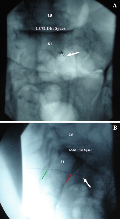
Fig. 3.
a AP image of needle in the right S1 foramen (white circle) of a cadaveric specimen. White arrow shows needle tip. b Lateral image depicting the needle penetrating into the pelvic cavity through the S1 ventral foramen. Green line delineating the dorsal sacral border. Red line delineating the ventral sacral border. White arrow illustrating the tip of the needle in the pelvic cavity of a cadaveric specimen

