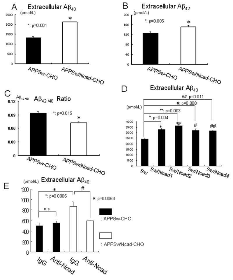
Figure 2. N-cadherin expression enhances extracellular Aβlevels and reduces Aβ42/40 ratio.

(A) APPSw-CHO cells or APPSw/Ncad-CHO cells were incubated in OPTI-MEM for 12 hours. The amount of extracellular Aβ40 was significantly elevated in APPSw/Ncad-CHO cells, compared to APPSw-CHO cells (n=8, p=0.001).
(B) APPSw-CHO cells or APPSw/Ncad-CHO cells were incubated in OPTI-MEM for 12 hours. After incubation, culture medium was collected and the amount of extracellular Aβ42 was measured. Extracellular Aβ42 was significantly elevated in APPSw/Ncad-CHO cells, compared to APPSw-CHO cells (n=8, p=0.005).
(C) The Aβ42/40 ratio in the medium was significantly decreased in APPSw/Ncad-CHO cells, compared to APPSw-CHO cells (n=8, p=0.015).
(D) APPSw-CHO (Sw) cells or four independent stable cell lines of APPSw/Ncad-CHO cells (SwNcad1–4) were incubated in OPTI-MEM for 24 hours. After incubation, the amount of extracellular Aβ40 was measured. Secreted extracellular Aβ40 was significantly elevated in every APPSw/Ncad-CHO stable cell line (SwNcad1–4), compared to that in APPSw-CHO cells (Sw) (n=4).
(E) APPSw-CHO cells or APPSw/Ncad-CHO cells were incubated in fresh OPTI-MEM containing either control IgG or N-cadherin-neutralizing antibody for 6 hours. After incubation, the amount of extracellular Aβ40 was measured. N-cadherin-neutralizing antibody significantly reduced the extracellular Aβ40 release into the medium in APPSw/Ncad-CHO cells (p=0.03, n=4). Conversely, N-cadherin-neutralizing antibody had no effect on the extracellular Aβ40 release into the medium in APPSw -CHO cells (p=0.3, n=4).
