
Figure 5.
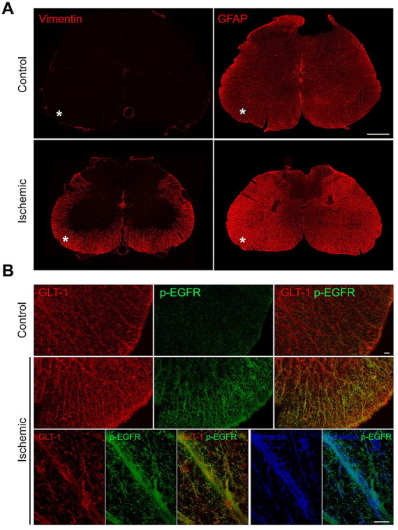
The EGF receptor is activated in vivo in the injured spinal cord. A, The increased vimentin and glial fibrillary acidic protein (GFAP) immunoreactivity reveals the presence of reactive astrocytes in the white matter of the lumbar spinal cord (asterisks) following an ischemic injury. Scale bar, 500 μm. B, Immunolabeling for phosphorylated EGF receptor (green) is increased in the white matter of the spinal cord after ischemia and shows substantial colocalization with the astrocytic glutamate transporter GLT-1 (red) as well as partial colocalization with the cytoskeletal protein vimentin (blue) in triple-labeled sections. Scale bars, 20 μm.