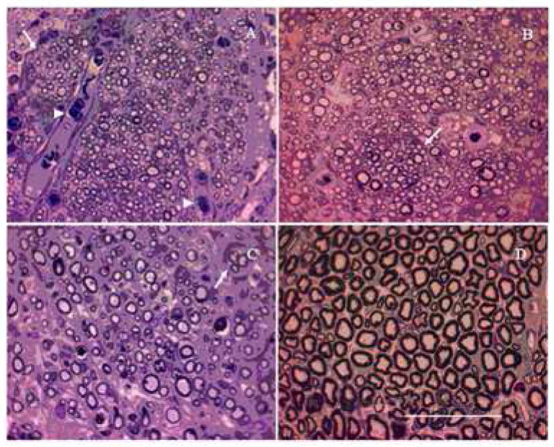
Figure 6.

Light micrographs of nerve sections from groups with successful axonal regeneration. Toludine-blue stained cross-sections at mid-length (10 mm) are shown here. Anisotropic scaffolds with step-gradient (A) and continuous-gradient (B) of LN-1, have higher density of axons and smaller-diameter axons than nerve grafts (C) and normal nerve (D). Note the fiber grouping in small fascicles (indicated by arrow) and presence of red blood cells (arrowhead). Scale bar = 50 μm
