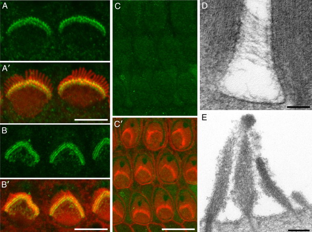
Figure 2.
Distribution of Vlgr1 in the mouse cochlea. A–C, Confocal images of sensory hair bundles of inner (A) and outer (B, C) hair cells from heterozygous (A, B) and homozygous (C) Vlgr1/del7TM mice double labeled with affinity-purified rabbit anti-mouse Vlgr1 antibodies (green) and Texas Red phalloidin (red). Note how Vlgr1 is distributed in a tight band around the base of the hair bundle (A, B). Hair bundle staining is not observed in the homozygous mutant (C). Scale bars: A, B, 5 μm; C, 10 μm. D, Transmission electron micrograph from a tannic acid-stained, wild-type mouse cochlear culture (P2 plus 1 d in vitro) showing ankle links interconnecting the stereocilia around the basal region of an outer hair cell bundle. Scale bar, 100 nm. E, Transmission electron micrograph of an outer hair cell bundle from a wild-type mouse cochlear culture (P1 plus 1 d in vitro) immunogold labeled with antibodies to Vlgr1. Gold particles are concentrated around the base of the hair bundle in the ankle link region. Scale bar, 200 nm.

