
Figure 6.
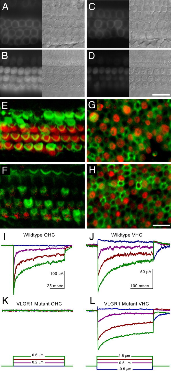
FM1-43 dye loading and transduction currents in Vlgr1/del7TM mutant mice. FM1-43 dye loading (A–D) of hair cells in cochlear cultures prepared from the apical (A, C) and basal (B, D) coils of heterozygous (A, B) and homozygous (C, D) Vlgr1/del7TM mice. Nomarski interference contrast images of the cells are shown adjacent to A–D. Images in A and C are focused at the level of the nucleus and were captured 6 min after the dye dip, and images in B and D are focused at the level of the hair bundle and were captured 5 min after the dye dip. Scale bar, 10 μm. Confocal images of FM1-43FX uptake (red) and phalloidin staining (green) in control (E, G) and Vlgr1/del7TM mutant (F, H) mouse hair cells at P7 from sensory cell epithelia excised from the apical turn of the cochlea (E, F) and from the utricle (G, H). Scale bar, 10 μm. Transduction current recordings in control (I, J) and Vlgr1/del7TM mutant (K, L) mouse hair cells at P7 from apical turn OHCs of the cochlea (I, K) and from vestibular hair cells (VHC) acquired from the utricle (J, L). Calibration in I and J apply to K and L, respectively.