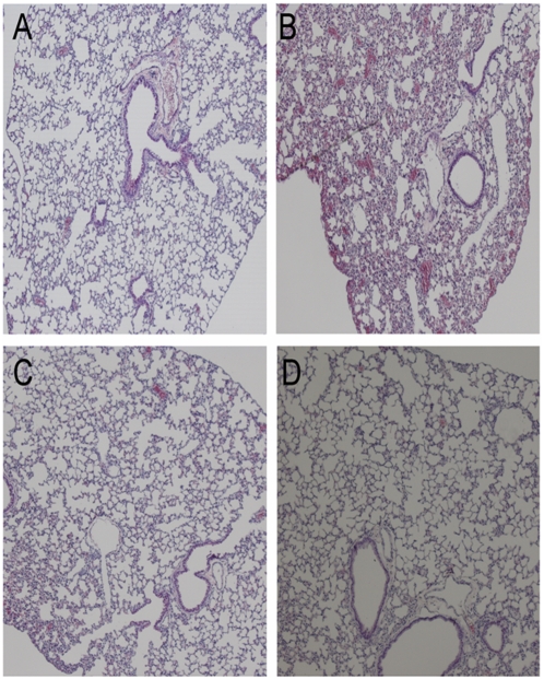
Figure 4. Histopathology of lung tissue in passively treated mice.
Photomicrographs of hematoxylin and eosin stained lung sections of mice treated with single or double doses of the combination of mAbs after post experimental viral infection with Clade 1 A/Vietnam/1203/2004 H5N1 virus at 6 days post challenge. A) Normal morphology seen in uninfected mice, B) infected and untreated mice, C) mice treated with a single dose of 5 mg/kg of ch-mAbs at 24 h post infection, D) mice treated with two doses of 5 mg/kg of ch-mAbs at 24 h and 72 h post infection.

