Figure 2.
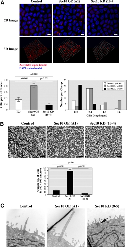
Sec10 knockdown leads to decreased primary ciliogenesis. (A) Control, Sec10-overexpressing (OE), and Sec10 knockdown (KD) MDCK cells were grown on Transwell filters for 14 d. Using antibody against acetylated α-tubulin that is specific for primary cilia, we examined ciliogenesis by immunofluorescence staining and confocal microscopy, as well as 3D reconstruction of the stacked series. Primary cilia were more abundant (as determined by the ratio of primary cilia/cell nuclei) and longer in Sec10 OE cells compared with control cells and less abundant and shorter in Sec10 KD cells compared with control cells. Quantification of ciliary abundance and cilia length are shown in the graphs below the images. Bar, 5 μm. The images are representative of >50 fields examined, with each experiment being repeated three times. Although MDCK cells are well ciliated and have long been used to study ciliogenesis (Praetorius and Spring, 2001), for unknown reasons cilia formation in MDCK cells is relatively sporadic, with the literature describing from ∼30% of normal MDCK cells being ciliated, as we see, to as little as 14.6% (Shalom et al., 2008). (B) To examine ciliogenesis by another method and in more detail, we grew control, Sec10 OE, and Sec10 KD cells on Transwell filters for 14 d. The cells were fixed in glutaraldehyde and SEM was performed (FEI Company XL20 SEM). A 200 mesh thinbar EM grid was placed on the samples and the number of cilia in a 100- × 100-μm area was determined. Confirming the results in Figure 2A, primary cilia seemed longer and therefore easily identifiable, in Sec10 OE cells compared with control cells and were rarely identified in Sec10 KD cells. Quantification of these results is shown on the graph below the images. Bar, 0.5 μm. (C) Using TEM, images of the cilia and basal bodies were occasionally captured. Sec10 KD cells demonstrated only basal bodies (arrow). In contrast, the control and Sec10 OE cells showed both basal bodies and elongated cilia, with the length of the cilia greater in the Sec10 OE cells. Bar, 0.5 μm.

