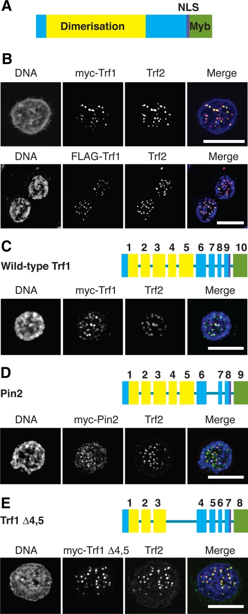
Figure 6.
Microscopy analysis of splice variants of Trf1. (A) Domain structure of chicken Trf1, based on published data for the human orthologue. (B) Wild-type DT40 cells were transfected with expression vectors for myc-Trf1 or FLAG-Trf1 and then fixed and stained with antibodies to myc or FLAG, respectively (green), and to Trf2 (red) and counterstained with DAPI (blue). Scale bars, 10 μm. Trf1−/−/− DT40 cells were transfected with expression vectors for myc-Trf1 (C), myc-Pin2 (D), or myc-Trf1Δ4,5 (E) and then fixed and stained with antibodies to myc (green) and Trf2 (red) and counterstained with DAPI (blue). Exon diagrams use the same color scheme as A and indicate the exons deleted in the splice variants of Trf1. Scale bars, 10 μm.

