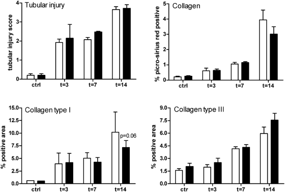
Figure 4. Analysis of UUO-induced tubular injury and fibrosis.
Tubular injury was semi-quantitatively scored using PAS-D stained renal tissue sections. Tubular damage was similar between TLR2+/+ (□) and TLR2−/− (▪) mice at all time points. Fibrosis was quantitatively scored by measuring renal collagen accumulation in TLR2+/+ and TLR2−/− mice 3, 7, and 14 days after UUO or in contralateral kidneys. This revealed that fibrosis was comparable in kidney of TLR2−/− mice compared with TLR2+/+ animals during UUO-induced injury. The percentage of positive staining for collagen (picrosirius red staining), collagen type I and III was analyzed in both cortex and medulla using a computer-assisted digital analysis program. Data are mean and SEM of six mice per group, *p<0.05.

