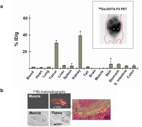
Figure 3. (a) Biodistribution of 213Bi-DTPA-[F3]2.
3.7 MBq of 213Bi-DTPA-[F3]2 were injected i.p. into mice bearing intra-peritoneal MDA-MB-435 xenograft tumors. After 45 minutes the 213Bi-DTPA-[F3]2 activity present in individual organs, the tumors and the blood was measured. Values represent the percentage of the injected dose/g tissue (%ID/g)±SEM. The inset depicts a mouse with peritoneal carcinomatosis imaged with 68Ga-DOTA-F3-PET. The asterics (*) indicates the location of the kidneys. b) Autoradiography studies were performed using histological sections of MDA-MB-435 xenograft tumors or muscle tissue. 213Bi-DTPA-[F3]2 was found in tumors 45 minutes after i. p. injection. 213Bi-DTPA-[F3]2 was found in the periphery of the tumor as well as in spots within the tumor tissue. H&E-staining and microscopy (100fold magnification) of the sections revealed that the intra-tumoral accumulation of 213Bi-DTPA-[F3]2 occurs in the perivascular region. The H&E-stained picture represents the area within the white box in the autoradiography picture. Negligible activities were found in autoradiography pictures of control organs such as muscle.

