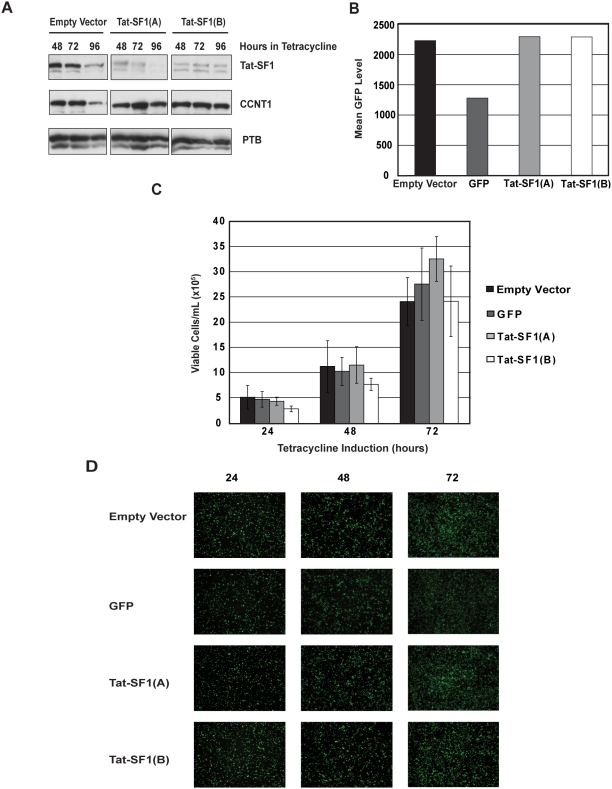
Figure 1. Tat-SF1 depletion does not affect T-Rex-293 cell viability.
(A) Analysis of Tat-SF1 knockdown by Western blot. T-Rex-293 cells were induced with tetracycline and lysates were made at the time points indicated. Equal protein amounts were loaded for Western blot analysis with antibodies against Tat-SF1, CCNT1 and PTB. Lanes 1–3 are from cells that express an empty vector control and lanes 4–9 are from cells that express one of two unique shRNAs targeting Tat-SF1. (B) Analysis of GFP knockdown by flow cytometry. T-Rex-293 cells were induced with tetracycline for 72 hours, and resuspended cells were fixed with formaldehyde for flow cytometry analysis. The bar graph quantifies the mean GFP level of each sample. (C) Analysis of cell viability by trypan blue exclusion. Equivalent numbers of T-Rex-293 cells (expressing either an empty vector control, an shRNA targeting GFP, or one of two shRNAs targeting Tat-SF1) were induced with tetracycline for the times indicated. Viable cells per mL are reported as a means of three independent experiments. Error bars represent standard error. (D) Analysis of cell viability by fluorescence microscopy. Equal numbers of T-Rex-293 cells were induced with tetracycline for the times indicated and imaged with fluorescence microscopy.

