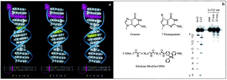
Figure 1.
The DNA assemblies. (a) Molecular models (Insight II) illustrating the E-tethered (red) DNA assemblies (left to right) 5Z, 6Z, and 7Z. The Z base is shown in yellow. Sequences are given below. (b) Structures of guanine (G), Z, and the E-modified tether, as well as an autoradiogram (right) after denaturing 18% PAGE, showing photoinduced damage of an E-modified duplex generated by irradiation at 313 nm of the duplex (10 μM) in 5 mM phosphate/50 mM NaCl, pH 7, 5′ 32P-labeled on the strand complementary to that containing the tether. Crosslinking occurs at the first two base steps on the 3′ side (near E); see The DNA Assemblies and ref. 35. The sequence 3′-CGCGCACTTA-5′ also was examined and gave consistent results.

