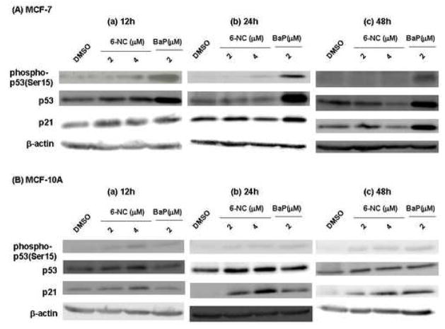
Fig. 2.
Representative Western blot analysis of phospho-p53(ser15), p53 and p21Cip1 proteins in MCF-7 cells (A) and MCF-10A (B) cells after exposure to 6-NC (2 μM and 4 μM) or BaP (2 μM) for 12, 24 and 48 h. Cells treated with DMSO alone were used as negative control. β-Actin was used as a loading control.

