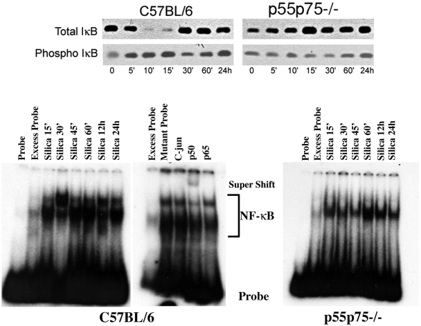
Figure 4. TNFα receptors mediate NF-κB activation in experimental silicosis.
Western blot (upper panel) representing expression of total and phosphorylated IκB (phospho IκB) in the lungs of silica-sensitive C57BL/6, or double (p55/p75−/−) TNFα receptor deficient mice following the intratracheal instillation of silica particles as described in the text. Lower panel illustrates the DNA binding activity of NF-κB in crude nuclear extracts from whole lung isolated from C57BL/6, and double (p55/p75−/−) TNFα receptor deficient mice after silica exposure. Silica induces two bands: a lower band representing the p50-p50 homo-dimer, present in both the C57BL/6 and the double (p55p75−/−) TNFα receptor deficient mice, and an upper band, only observed in the lungs of C57BL/6 mice, representing the p50-p60 heterodimer. Excess probe represents NF-κB binding in lung nuclear extract of a silica-treated mouse, assayed in the presence of excess unlabelled oligonucleotide as a competitor. Competition assays were performed using 400 times excess of unlabeled probe or NF-κB mutant oligonucleotide (Santa Cruz). Supershifts were performed by adding to the binding mixture antibodies to p50 (sc1190X) or to p65 (sc372X) or against C-Jun/AP-1 (sc44X) (Santa Cruz) before adding the labeled probe to the mixture.

