Figure 1.


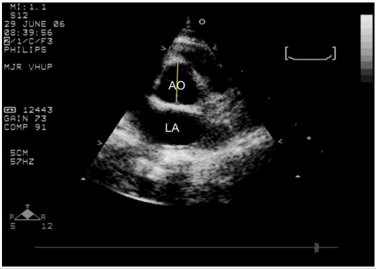
- A short axis right parasternal echocardiogram from an unaffected cat showing where the aortic diameter is measured at the level of the aortic valve (yellow line). AO= aorta; LA= left atrium.
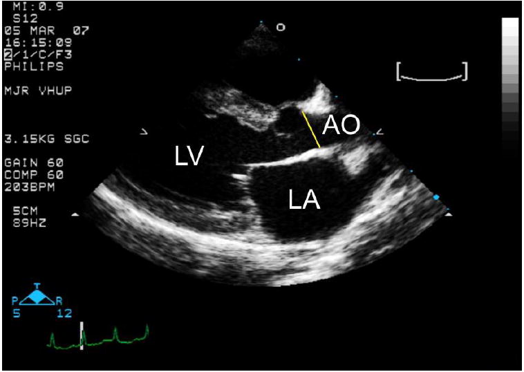
- A long axis right parasternal echocardiogram from an unaffected cat showing the sinotubular junction, the aortic region most frequently dilated in cats with MPS I and VI. AO= aorta; LA= left atrium.
