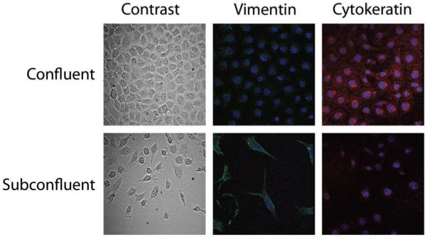
Figure 3. Mouse mesonephric M15 cells mimicking the mesenchymal to epithelial transition during nephrogenesis.

M15 cells grown at different cell densities showing an epithelial morphology (phase contrast microscopy) and cytokeratin expression (red immunofluorescence) at confluence, and a mesenchymal phenotype (spindle shaped morphology and vimentin expression, green) when grown in subconfluent conditions. Nuclei were counterstained with TOPRO-3.
