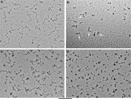
FIGURE 3.
Visualization of C-rich and G-rich telomeric transcripts. C-rich (A) and G-rich (B) RNA molecules in 100 mm KCl were prepared for EM by mounting on thin carbon supports, dehydrating, and rotary shadowcasting with tungsten. The C-rich RNA appears as an extended thread with kinks and compact regions. The G-rich RNA appears as a mixture of balls and thick rods (arrows). The thickness of the rods is significantly greater than that of the C-rich RNA or duplex DNA. C-rich (C) and G-rich (D) RNA molecules were mounted in 10 mm KCl as in A and B. The C-rich RNA again appears extended with kinks, whereas the G-rich RNA appears as mostly balls as opposed to a mixture of rods and balls. The bar is equivalent to 100 nm.

