FIGURE 6.
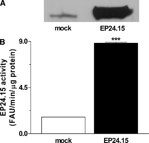
Quantification of EP24.15 overexpression in HEK293 cells in Experiment 2. HEK293 cells were transiently transfected with empty pShooter (mock-transfected cells) or with pShooter coding for EP24.15 (EP24.15-transfected cells). A, proteins from crude cell extract (62.5 μg from mock cells and 5 μg from cells transfected with EP24.15) were separated by SDS-PAGE on an 8% polyacrylamide gel and transferred to nitrocellulose membranes, which were incubated with specific rabbit antiserum against EP24.15 (1:3000). After incubation with an anti-rabbit IgG-horseradish peroxidase-conjugated secondary antibody (1:3000), the immunoreactive bands were visualized by chemiluminescence. B, EP24.15 enzymatic activity was determined in triplicate using a continuous assay with a quenched fluorescent substrate as described under “Experimental Procedures.” EP24.15 activity was about six times greater in EP24.15 overexpressing cells then in the mock control group. ***, p < 0.001, statistically different from mock control group using Student's t test.

