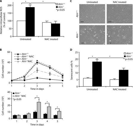
FIGURE 2.
Decreased proliferation in Atm-/- astrocytes is caused by elevated ROS levels. A, Atm+/+ and Atm-/- astrocytes were either left untreated or pretreated with NAC (1 mm) for 2 h. Fluorescent H2DCFDA levels were determined for these cells. The means ± S.D. of three independent experiments are shown. *, p < 0.05 when untreated Atm-/- culture were compared with untreated Atm+/+ astrocytes or when NAC-treated Atm-/- astrocytes were compared with untreated Atm-/- astrocytes. B, growth curves of Atm-/- astrocytes in the presence or absence of NAC (1 mm) were analyzed for 5 days and then compared with those of Atm+/+ astrocytes. The means ± S.D. of three independent counting are shown. *, p < 0.05 when NAC-treated Atm-/- astrocytes were compared with untreated Atm-/- astrocytes. C, photomicrographs of Atm+/+ and Atm-/- astrocytes in the presence or absence of NAC. Scale bars, 50 μm. D, senescence of Atm+/+ and Atm-/- astrocytes in the presence or absence of NAC was compared by SA β-galactosidase expression 2 days after cultivation. The means ± S.D. of three independent experiments are shown. *, p < 0.05 when untreated Atm-/- culture was compared with untreated Atm+/+ astrocytes or when NAC-treated Atm-/- astrocytes was compared with untreated Atm-/- astrocytes.

