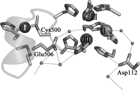
FIGURE 2.
Structure around the active site of the truncated mutant of CueO (7). Type I, II, and III coppers are represented as spheres. Small spheres, oxygen atoms. The two networks of hydrogen bonds lead to the exterior of the protein molecule, forming the pathway to let protons in and water molecules out. Mutated amino acid residues, Cys500, Glu506, and Asp112, and the networks of hydrogen bonds are indicated.

