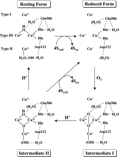
FIGURE 6.
The four-electron reduction of dioxygen by CueO. The structure of
the resting CueO is based on the x-ray crystal structure of rCueO
(5) and the truncated mutants
(7). It is not known whether
water molecules are present near the active site in the reduced CueO. The most
probable peroxide-bound structure for intermediate I is figured to account for
the production of the charge transfer bands due to
 → Cu2+ and to be
EPR-silent. Intermediate I is converted into intermediate II with the supply
of electrons from type I and II coppers and of a proton with the assistance of
Glu506. In intermediate II, four electrons are transferred to
dioxygen, and the O–O bond is cleaved. Type II and III coppers are
magnetically interacted to give the g < 2 EPR signal. In the decay
of intermediate II, another proton is donated with the assistance of
Glu506.

