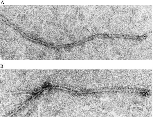
Figure 5.
Electron micrographs showing antigen-specific labeling of filamentous phage displaying the 2H6 Fv heterodimer. Similar results were obtained for the 21H3 phage Fv; however, the micrographs were not as distinct. (A) A phage specifically labeled with a 5-nm colloidal gold particle adhered to the PCP-BSA antigen (×105,000). (B) A phage on the same grid as in A labeled with two gold particles (×105,000).

